Nstemi Ecg - Accuracy Of Omi Ecg Findings Versus Stemi Criteria For Diagnosis Of Acute Coronary Occlusion Myocardial Infarction Sciencedirect / Type 2 has been reported up to 25% of cases of mi depending on the population studied.
Nstemi Ecg - Accuracy Of Omi Ecg Findings Versus Stemi Criteria For Diagnosis Of Acute Coronary Occlusion Myocardial Infarction Sciencedirect / Type 2 has been reported up to 25% of cases of mi depending on the population studied.. Where to look on an ecg for stemi and nstemi the beauty of ecg changes consistent with myocardial ischaemia, injury and infarction is that they all show up differently on a 12 lead ecg. As discussed previously, ischemia results in ecg changes. The ecg tracing can have. Chest pain is the second most common complaint. Unstable angina and nstemi differ primarily in the presence or absence of detectable troponin leak.
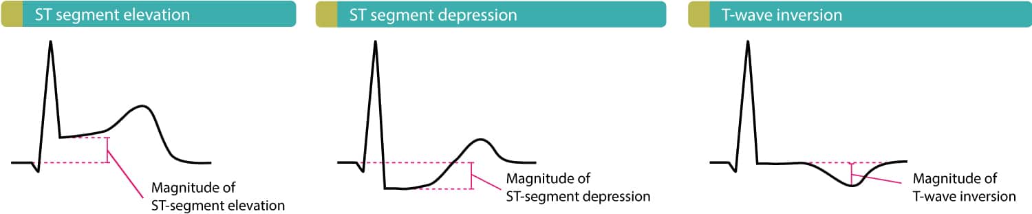
Unstable angina and nstemi differ primarily in the presence or absence of detectable troponin leak. Diagnostic values of combined ecg findings for differentiating ttc from nstemi in the setting of non st‐elevation ecg are shown in table 4. Nstemi is diagnosed in patients determined to have symptoms consistent with acs and troponin elevation but without ecg changes consistent with stemi. T‐inversion in more than 5 of any leads had a sensitivity of 36% to diagnose ttc in this particular setting with a specificity of 92% ( p <0.001, with ppv of 80% and npv of 60%). But before we talk about these changes, we should quickly brush up on what the normal components of an ecg trace look like:
Cureus Frequency Of Non St Segment Elevation Myocardial Infarction Nstemi In Acute Coronary Syndrome With Normal Electrocardiogram Ecg Insights From A Cardiology Hospital In Pakistan from assets.cureus.com 1/3 will have stemi, 2/3 nstemi. This ecg is reproduced from an article by zajarias et al. Type 2 mi is defined as myocardial infarction secondary to ischaemia due to either increased oxygen demand or decreased supply, e.g. Stemi results from complete and prolonged occlusion of an epicardial coronary blood vessel and is defined based on ecg criteria.nstemi usually results from severe coronary artery narrowing, transient occlusion, or microembolization of thrombus and/or atheromatous material. A myocardial infarction is the medical term for a heart attack. Ecg changes, pathological q waves, or imaging evidence of new loss of viable myocardium or new regional wall motion abnormality. T‐inversion in more than 5 of any leads had a sensitivity of 36% to diagnose ttc in this particular setting with a specificity of 92% ( p <0.001, with ppv of 80% and npv of 60%). Nstemi is diagnosed through a blood test and an ecg.
T‐inversion in more than 5 of any leads had a sensitivity of 36% to diagnose ttc in this particular setting with a specificity of 92% ( p <0.001, with ppv of 80% and npv of 60%).
Aims recognise the ecg patterns which occur in nstemi focus on those which occur most commonly difficult ecg scenarios. However, it may also be normal or show nonspecific changes. Acute coronary syndrome is caused by a mismatch between myocardial oxygen demand and myocardial oxygen delivery. Type 2 has been reported up to 25% of cases of mi depending on the population studied. Coronary artery spasm, coronary embolism, anaemia, arrhythmias, hypertension or hypotension. the definition of type 2 mi is unsatisfactory because it is not really defined by what it is but rather what it is not. Nstemi is diagnosed in patients determined to have symptoms consistent with acs and troponin elevation but without ecg changes consistent with stemi. In fact, the type of ischemia will determine which type of ecg changes that occur. An electrocardiogram or ecg that displays each heartbeat as a waveform is. Unstable angina and nstemi differ primarily in the presence or absence of detectable troponin leak. This pattern is consistent with an acute infarction localised to the superior portion of the lateral wall of the left ventricle (high lateral stemi). Where to look on an ecg for stemi and nstemi the beauty of ecg changes consistent with myocardial ischaemia, injury and infarction is that they all show up differently on a 12 lead ecg. Ecg (ekg) in acute stemi (st elevation myocardial infarction) the ecg is the key to diagnose stemi. An acute coronary syndrome may include various clinical entities that involve some sort of ischemia or infarction.
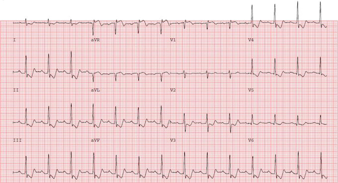
Nstemis are a type of acute coronary syndrome and are defined by the presence of myocardial infarction as detected by a rise in cardiac biomarkers, without ecg changes indicative of a stemi. Page one contains the limb leads, and page two shows us the precordial leads. The culprit vessel in this case was an occluded first diagonal branch of the lad. Chest pain is the second most common complaint. However, it may also be normal or show nonspecific changes.
Classification Of Acute Coronary Syndromes Acs Acute Myocardial Infarction Ami Ecg Echo from ecgwaves.com However, it may also be normal or show nonspecific changes. Where to look on an ecg for stemi and nstemi the beauty of ecg changes consistent with myocardial ischaemia, injury and infarction is that they all show up differently on a 12 lead ecg. The rhythm is atrial fibrillation, with a heart rate of 133 bpm and an. Ecg changes, pathological q waves, or imaging evidence of new loss of viable myocardium or new regional wall motion abnormality. A myocardial infarction is the medical term for a heart attack. In the stemi paradigm of acute myocardial infarction (ami), many nstemi patients have unrecognized acute coronary occlusion mi (omi), may not receive emergent reperfusion, and have higher mortality than nstemi patients without occlusion. This ecg is reproduced from an article by zajarias et al. Diagnostic values of combined ecg findings for differentiating ttc from nstemi in the setting of non st‐elevation ecg are shown in table 4.
Type 2 mi is defined as myocardial infarction secondary to ischaemia due to either increased oxygen demand or decreased supply, e.g.
In the journal of invasive cardiology. Aims recognise the ecg patterns which occur in nstemi focus on those which occur most commonly difficult ecg scenarios. In the stemi paradigm of acute myocardial infarction (ami), many nstemi patients have unrecognized acute coronary occlusion mi (omi), may not receive emergent reperfusion, and have higher mortality than nstemi patients without occlusion. The culprit vessel in this case was an occluded first diagonal branch of the lad. Nstemi is diagnosed in patients determined to have symptoms consistent with acs and troponin elevation but without ecg changes consistent with stemi. Ecg changes, pathological q waves, or imaging evidence of new loss of viable myocardium or new regional wall motion abnormality. Time is muscle when treating heart attacks. Acute myocardial infarction (mi) significantly contributes to mortality and morbidity in developed and developing countries. As discussed previously, ischemia results in ecg changes. Type 2 has been reported up to 25% of cases of mi depending on the population studied. 25% will be diagnosed with acs. This pattern is consistent with an acute infarction localised to the superior portion of the lateral wall of the left ventricle (high lateral stemi). Acute coronary syndrome is caused by a mismatch between myocardial oxygen demand and myocardial oxygen delivery.
Acute myocardial infarction (mi) significantly contributes to mortality and morbidity in developed and developing countries. The rhythm is atrial fibrillation, with a heart rate of 133 bpm and an. Ecg diagnostics for myocardial infarction. Type 2 has been reported up to 25% of cases of mi depending on the population studied. Coronary artery spasm, coronary embolism, anaemia, arrhythmias, hypertension or hypotension. the definition of type 2 mi is unsatisfactory because it is not really defined by what it is but rather what it is not.
Non St Elevation Myocardial Infarction Diagnosis And Management Intechopen from www.intechopen.com 1/3 will have stemi, 2/3 nstemi. Aims recognise the ecg patterns which occur in nstemi focus on those which occur most commonly difficult ecg scenarios. But before we talk about these changes, we should quickly brush up on what the normal components of an ecg trace look like: The ecg tracing can have. Diagnostic values of combined ecg findings for differentiating ttc from nstemi in the setting of non st‐elevation ecg are shown in table 4. An acute coronary syndrome may include various clinical entities that involve some sort of ischemia or infarction. Stemi results from complete and prolonged occlusion of an epicardial coronary blood vessel and is defined based on ecg criteria.nstemi usually results from severe coronary artery narrowing, transient occlusion, or microembolization of thrombus and/or atheromatous material. Pain compatible with angina pectoris and.
Pain compatible with angina pectoris and.
These markers are evidence of. Ecg which does not meet the criteria for stemi or stemi equivalent and. An nstemi differs from a stemi, which is the most common type of heart attack, by causing less damage to a person's heart. Ecg diagnostics for myocardial infarction. 1/3 will have stemi, 2/3 nstemi. This pattern is consistent with an acute infarction localised to the superior portion of the lateral wall of the left ventricle (high lateral stemi). Unstable angina and nstemi differ primarily in the presence or absence of detectable troponin leak. We have proposed a new omi vs. 25% will be diagnosed with acs. In the journal of invasive cardiology. Type 2 mi is defined as myocardial infarction secondary to ischaemia due to either increased oxygen demand or decreased supply, e.g. Learn how to diagnose st elevation myocardial infarction on ecg and how to determine the location of the infarct. But before we talk about these changes, we should quickly brush up on what the normal components of an ecg trace look like:
Ecg which does not meet the criteria for stemi or stemi equivalent and מדא. Rise or fall of cardiac biomarker values with at least one value above the 99th percentile, possibly by point of care measurement and/or.


0 Komentar